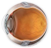
lens
Removing cataract and implantation of an artificial lens doesn’t differ that much from such procedures in people who don’t have Wagner. It is preferable that such a procedure is done by an experienced surgeon in connection to the abnormal vitreous and possible complications like retinal detachment.
A complication that occurs more frequently is the onset of an epiretinal membrane, that starts growing on the retina. If this membrane disturbs vision, or when it causes traction on the retina and there is a obvious risk on tearing or detachment, the membrane can be removed in a procedure called a vitrectomy (see below).
retina
Retinal breaks, without fluid pushing itself underneath it, can be treated with laser or cryocoagulation. The purpose is to seal the retina around the break on the underlying layer to prevent larger breaks or retinal detachment.
When retinal detachment occurs, when fluid is underneath it, incisional surgery is needed. The first method is scleral buckling. The surgeon will put a silicone band around the eyeball, making this somewhat smaller, relieving the tension on the retina.
treatments
This kind of procedures can lead to the onset of cataract, also in the fellow eye.
The succesrate of procedures for reattaching the retina in people with Wagner is not so favourable.
prophylactic procedures
The retina in people with Wagner is fragile. Parts can be affected by lattice degeneration, mainly in the peripheral retina with the risk of tearing of the retina. Laser seems to be ineffective in Wagner patients, in young <15 years of age), highly mopic Wagners it can cause retinal detachment. Maybe scleral buckling is a better option.
VCAN-related vitreoretinopathy is inherited in an autosomal dominant manner. Most individuals diagnosed with VCAN-related vitreoretinopathy have an affected parent. Each child of an affected individual has a 50% chance of inheriting the mutation.
Although no laboratories listed in the GeneTests Laboratory Directory offer molecular genetic testing for prenatal diagnosis of VCAN-related vitreoretino-pathy, such testing may be possible through laboratories offering custom prenatal testing for families with a known disease-causing mutation.
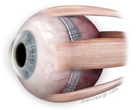
fig. Scleral buckling: a silicon band around the eyeball relieves the pressure on the retina
The second method is a vitrectomy. The vitreous is removed and the tears sealed. The vitreous is replaced with gas or oil. The gas dissappears by itself, oil has to be removed in a second operations. The vitreous cavity if filled with aqueous humour, produced by the ciliary body in the front of the eye. Sometimes this procedure is accompanied by scleral buckling to relieve the tension on the retina.
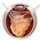
fig. Vitrectomy: the vitreous is being removed. 3 tubes are being used: a drip to stabilise the quantity of fluid -and the intraocular pressure- inside the eye, a lamp and a piston. Right: complete detached retina with instruments.
Illustrations: with courtesy of www.jirehdesign.com
surgical techniques
With incisional surgery the conjunctiva are opened and the healing will be accompanied by scarring. This makes other procedures in the future more difficult. So some surgeons take this into account and are using techniques that are not compromising the conjunctiva.
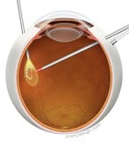
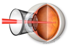
fig. Laser photo-coagulation seals the retina on the layers underneath
page last modified Mar 30th 2012
Wagner syndrome