pedigrees
the Netherlands
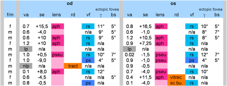
Jansen 1 (1966)
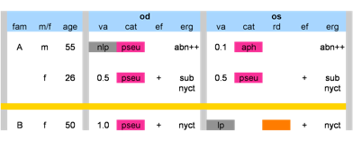
The first person identified with Wagner in the Netherlands is a woman who lived between 1840-1905 in NIjmegen. In the 1960s Jansen came across one of her great-grandchildren. In total he saw 39 affected family members of which he included 29 as family B In his thesis. The genetic variant was determined as c.4004-5T>C.
Jansen 2 (1966)
This pedigree was also included in Jansen’ thesis, and was indicated as family A with 4 affected members. Jansen didn’t exclude the possibility that the two families, as they both came from the same region, might be related in earlier generations.
the Netherlands
Mukhopadhyay (2006)
Jansen 1 (1966)
Jansen 2 (1966)
Switzerland
Kloeckener (2006)
Graemiger (1995)
France
Zech (1999)
Japan
Miyamoto (2005)
Great-Britain
Meredith (2007)
Fryer (1990)
USA
Ronan (2009)
ERVR
the Netherlands
Mukhopadhyay (2006)
USA
Brown (1995)
erosive vitreoretinopathy
USA
Brown (1994/95)
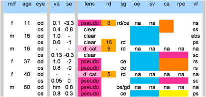
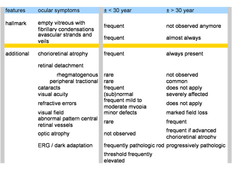
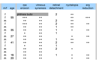
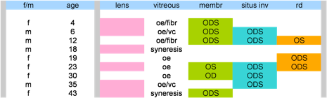
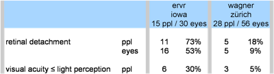
Brown et al from Iowa, USA described a pedigree with vitreoretinopathy, which he now categorised as a new clinical entity (1994) as the RPE initially seems normal, but later in life progressively thins or ‘erodes’ in the equatorial periphery. He describes 15 family members, 11 of them had rhegmatogenous retinal detachments, 6 eyes of 30 had a visual acuity that was reduced to light perception.
Switzerland
Graemiger (1995)
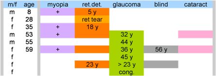
Graemiger evaluated the original pedigree described by Hans Wagner in 1938. He studied 28 family members. The most prominent finding was that retinal detachment indeed was part of the disorder. Wagner did not report retinal detachment in his article, but Jansen in the Netherlands did. So this finding made those entities more alike.
Brown (1995)
added about this pedigree that 5 people had a rhegmatogenous retinal detachment, 11 had bilateral peripheral tractional retinal detachments without retinal breaks and only 3 eyes had visual acuities of light perception or worse.
Kloeckener (2006)
Barbara Kloeckener et al confirmed that the ocular disorder in the Swiss pedigree is caused by the mutated Versican gene too: c.4004+1G>A. They concluded that there is a complete lack of exon 8 (see genetics). This shortens the predicted protein by 1754 amino acids and leads to severe reduction of glycosaminoglycan attachment sites. They tested 21 affected family members.
France
Zech (1999)
Zech identified 20 family members with Wagner disease.
Age range was 5-82, median 33 years.
Japan
Miyamoto (2005)
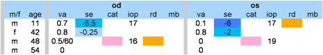
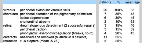
Miyamoto et al identified 11 family members (Tokushima Pedigree). They saw without exception an empty vitreous, avascular membrane and chorioretinal atrophy. The ERG was subnormal with everyone where assessment was done (2 eyes were not assessed (n/a), 2 were non-recordable).
In this family there was no elevated IOP nor glaucomatous damages.

Great-Britain
Meredith (2007)
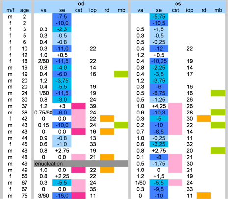
Meredith et al described 2 people with Wagner syndrome: one father and daughter and a third not related person (B) with Wagner features but not the mutated VCAN gene. The genetic variant in family A : c.9265+1G>A
All of them had the vitreous abnormalities: membranes, (de)pigmentation, and chorioretinal atrophy. With the oldest family member the intraocular pressure went out of control in both eyes after cataract surgery. Father and daughter from family A suffered from uveitis.
Mukhopadhyay (2006)
In the linkage study of Mukhapadyay et al eight families were involved: six families from Dutch and one of Chinese origin with Wagner disease, and one family with ERVR. 5 families, including the ERVR family had the founer mutation: c.4004-5T>C, two small families had respectively c.4004-5T>A en c.4004-1G>A. One of the Dutch Wagner families was also subject of the study of Jansen in the 60s. As this study was not focused on phenotype the clinical features are only summarily described.
The data of the ERVR-family you will find under de ervr-header at the bottom of this page.
the Netherlands
Mukhopadhyay (2006)
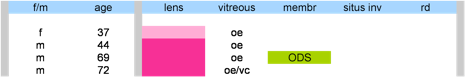
In the Netherlands a family wit ERVR was described in the linkage study on Wagner syndrome as they found that the same mutation in the Versican gene was the cause of this disorder too: c.4004-5T>C. So the two disorders are allelic. They described only a few family members.

page last modified Nov 18th 2009
Wagner syndrome
Fryer (1990)
Fryer et al decribed a Welsh family, of which was determined the diagnosis of Wagner syndrome by Graeme Black in Manchester in 1999 (family W1).
In this pedigree glaucoma was mentioned as one of the features, like in Jansen’s. We did not yet establish if this meant only an elevated IOP or also glaucomatous damage. Retinal detachment was a frequent finding in this pedigree.


Ronan (2009)
In Durham, North Carolina, USA a family was described: 6 people in 3 generations. THe genetic cause was found to be a mutation in the same base pair as the Zürich and Cambridg families, except the G was replaced by a T: c.9265+1G>T.